
An electrocardiogram (ECG) is a simple, non-invasive test that records the electrical activity of the heart. It is a crucial tool for monitoring heart function, detecting rhythm abnormalities, and guiding clinical decisions.
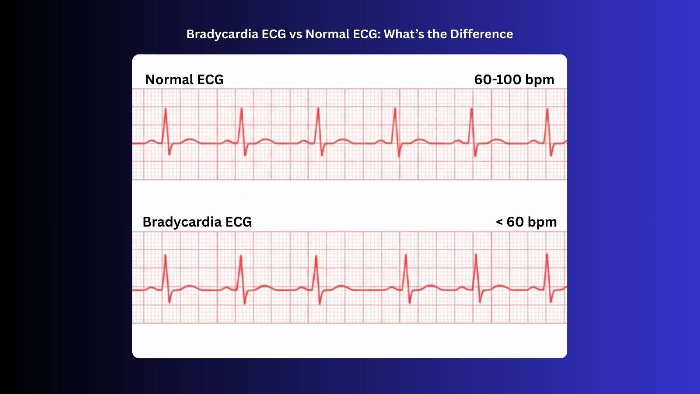
A normal heart rate in adults typically ranges from 60 to 100 beats per minute (bpm), reflecting a healthy and regular heart rhythm. In contrast, bradycardia is defined as a heart rate below 60 bpm, which may occur in healthy individuals, such as athletes, or as a result of underlying medical conditions. For those looking to deepen their skills, mastering ECG rhythms recognition and interpretation for ACLS is essential.
The purpose of this article is to clearly compare ECG characteristics in bradycardia versus a normal ECG, helping healthcare professionals, students, and enthusiasts understand how these differences appear on the heart’s electrical tracing and what they mean for patient care.
What is a Normal ECG
A normal ECG represents the heart’s electrical activity when it is functioning correctly. It provides insight into the heart rate, rhythm, and overall cardiac health and is generally identified as a Normal Sinus Rhythm (NSR). For a deeper look at each component of the ECG tracing, including the significance of the PQRST wave, see our guide to ECG interpretation. Key Features of a normal ECG include:
1. Heart Rate:
- Normal adult heart rate ranges from 60 to 100 bpm.
- The rhythm is typically regular, meaning the time between each heartbeat is consistent.
2. P Wave:
- Represents atrial depolarization, the electrical signal that triggers the atria to contract.
- Usually upright in most leads and uniform in shape.
3. PR Interval:
- The time from the start of the P wave to the start of the QRS complex.
- Normal duration: 0.12–0.20 seconds.
- Indicates the time it takes for the electrical signal to travel from the atria to the ventricles.
4. QRS Complex:
- Represents ventricular depolarization, which triggers ventricular contraction.
- Normal duration: 0.06–0.10 seconds.
5. T Wave:
- Represents ventricular repolarization, or the recovery phase of the ventricles.
- Usually upright in most leads.
6. QT Interval:
- From the start of the QRS complex to the end of the T wave.
- Normal duration varies with heart rate (generally 0.36–0.44 seconds).
- Important for assessing the risk of arrhythmias.
What is Bradycardia ECG
Bradycardia, defined as a heart rate below 60 beats per minute (bpm) in adults, is primarily regulated by the autonomic nervous system (ANS), which balances the parasympathetic division (vagus nerve), releasing acetylcholine to slow the heart, and the sympathetic division, releasing norepinephrine to speed it up; PNS dominance can cause bradycardia, especially during rest or relaxation. While a slower heart rate may be normal in some individuals, such as well-trained athletes, it can also signal underlying medical issues when accompanied by symptoms.
1. Causes of Bradycardia
Bradycardia happens when your heart beats slower than normal. It can show up naturally in some people or because of certain medical conditions.
1a. Physiological (Normal) Causes:
Some people naturally have a slower heartbeat and feel perfectly healthy. This can happen when the body is strong, relaxed, or well-trained.
- High Physical Fitness or Athletic Conditioning: People who exercise a lot or train their bodies well often have slower heartbeats because their heart works more efficiently. A strong heart doesn’t need to beat as fast to keep the body moving.
- Sleep or Relaxation: When you rest or sleep, your heart naturally slows down to save energy. This slower pace helps your body recharge and feel refreshed.
- Certain Medications, Like Beta-Blockers: Some medicines make your heart beat slower on purpose to protect it. These drugs help control blood pressure and reduce stress on the heart.
1b. Pathological (Medical) Causes:
Sometimes a slow heartbeat happens because of health problems. These medical issues can affect how the heart works and make it beat slower than normal.
- Heart Conditions, Such as Sick Sinus Syndrome or Heart Block: Some heart problems make the heartbeat slow because the heart’s natural rhythm gets interrupted. This can make you feel tired or dizzy.
- Hypothyroidism: When your thyroid gland works too slowly, it can slow down your heartbeat. Your body may also feel sluggish or low on energy.
- Electrolyte Imbalances: Having too much or too little of certain minerals in your blood can affect your heart rate. This can make your heartbeat irregular or unusually slow.
- Myocardial infarction (heart attack): A heart attack can damage the heart’s electrical system and slow your pulse. This can be serious and needs quick medical attention.
2. Symptoms of Bradycardia
A slow heartbeat can cause different feelings in your body. You might notice tiredness, dizziness, or trouble catching your breath.
- Fatigue or Weakness: A slow heartbeat can make you feel unusually tired or weak. Even small tasks may feel harder than usual.
- Dizziness or Lightheadedness: When your heart beats slowly, your brain might not get enough blood, making you feel dizzy. You may feel unsteady or woozy.
- Shortness of Breath: A slow heart can make it harder for your body to get enough oxygen. You might notice it’s tough to catch your breath.
- Fainting or Near-Fainting Episodes: Bradycardia can sometimes cause you to faint or feel like you might pass out. It happens when your brain doesn’t get enough blood for a moment.
3. Clinical Significance
- In many cases, bradycardia is benign and does not require treatment.
- However, when bradycardia causes symptoms or is due to heart disease, it can be dangerous, requiring medical evaluation and sometimes interventions like a pacemaker.
4. ECG Clues:
- On an ECG, bradycardia is primarily seen as fewer QRS complexes per minute.
- The rhythm may remain regular (sinus bradycardia) or show irregularities in AV (Atrioventricular) blocks.
Normal ECG vs Bradycardia
Bradycardia is primarily defined by a slower heart rate, but it also produces distinct patterns on the ECG compared to a normal heart rhythm. Understanding these differences is essential for accurate diagnosis.
| Feature | Normal ECG | Bradycardia ECG |
| Heart Rate | 60–100 bpm | <60 bpm |
| Rhythm | Regular | Usually regular, can be irregular depending on the cause |
| P Wave | Present before each QRS | Present before each QRS (can be normal or altered in some AV blocks) |
| PR Interval | 0.12–0.20 sec | Usually normal (0.12–0.20 sec) unless an AV block is present |
| QRS Complex | <0.12 sec | Usually normal (<0.12 sec); widened with bundle branch block. |
| T Wave | Upright in most leads | Usually normal; peaked or inverted with ischemia or electrolyte abnormalities. |
| R-R Interval | Consistent | Prolonged (longer than normal) |
| Clinical Significance | Normal cardiac function | Normal in athletes or during sleep; suggests sinus node dysfunction, AV block, hypothyroidism, or drug effects. |
Spotting the Signs: Normal vs Slow Heartbeats
In short, understanding the differences between a normal ECG and a bradycardia ECG helps you see how the heart’s electrical activity reflects its health. While a normal ECG shows a steady, regular rhythm with expected wave patterns, bradycardia slows the heartbeat and can change the timing of the electrical signals. Recognizing these patterns is important for spotting potential issues early and deciding when medical attention is needed. With practice, reading ECGs becomes easier, and knowing what to look for makes caring for patients safer and more confident.
Want to be ready for any emergency? Bayside CPR provides streamlined, flexible lifesaving training designed for busy schedules. Complete a short online course followed by a 30-minute in-person skills session at any of our 60+ locations to earn your AHA course completion card in ACLS, BLS, PALS, CPR, or First Aid. You’ll leave the same day prepared to act with confidence when it matters most.